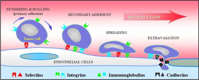
Fig. 1 Interactions between circulating cells and the endothelium
Mechanical forces at various scales (AFM Platform)
Micro-nanomechanical forces (Fig. 2) are measured when studying cellular systems (see review paper in J. Theoretical Medicine).
This approach aims at measuring two types of properties: cell morphology and microrheology (topography, elasticity maps), cell-cell adhesion measurements (receptor-ligand bonds)
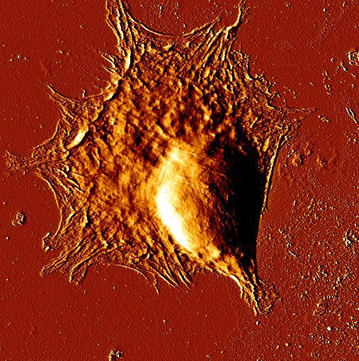
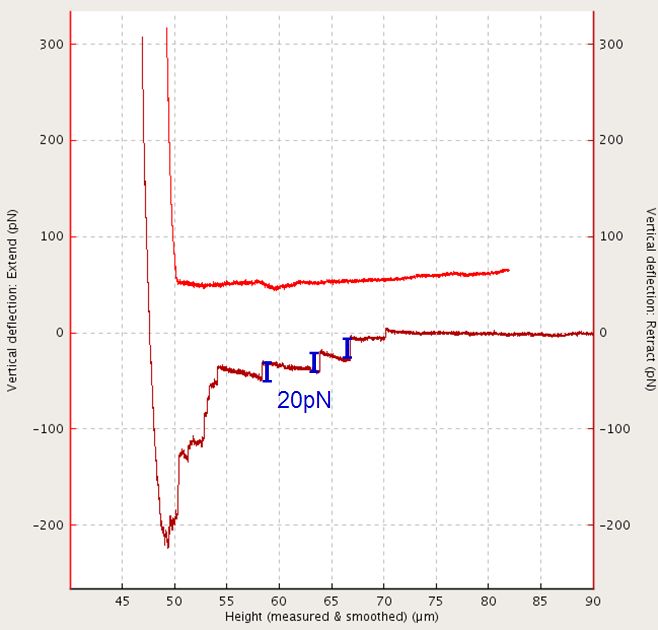
Fig. 2 Nanorheology (JPK AFM on inverted Zeiss microscope) - Topography of a fibroblast - Cell-cell separation and related force peaks (20pN each)
Another technique has been developped, based on indentation with superposed oscillations, leading to viscoelastic properties (Fig. 3). These properties have been studied on gels of different elasticity and on an endothelium layer. The results obtained show mechanosensitivity of cancer cells on these different substrates.
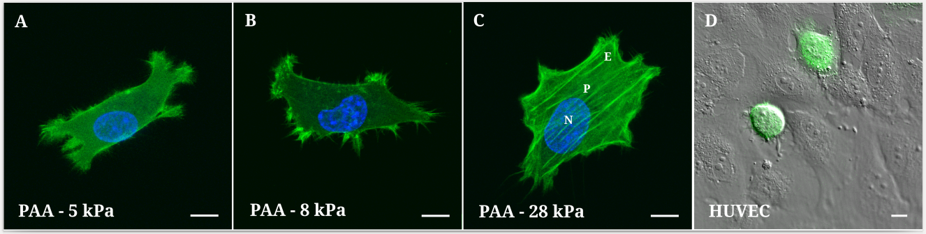
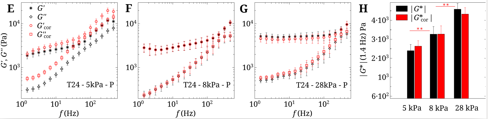
Fig. 3 Top: Cancer cells on different substrates (3 gels+endothelium). Bottom: Viscoelastic moduli (Pa)
Interactions between cells and the endothelium under flow
Mechanisms governing the interactions between cells and the endothelium have been studied using a flow chamber (closed circuit, with pressure and flow rate associated measurements) or microchannels (made at LIPhy) where fluids are moved using syringues pumps. Two situations were studied : the rolling of leucocytes (Fig. 4a), and the extravasation of cancer cells (Fig. 4b).
Fig. 4a Leukocyte rolling on the endothelium Fig. 4b Tumor cell extravasation
Cell migration - Traction forces
During many processes (migration, embryogenesis, wound healing, etc. ) cells have the ability to migrate in a certain environment. This work has enabled the determination of traction forces exerted by cells on 2D substrates. The images below show the different steps of the mesenchymal migration scheme (Fig. 5) : 1) lamellipodium extension 2) formation of adhesion sites and traction 3) contraction of the cell body d) release of the uropod
At the same time, we studied the effect of rigidity of the substrate. This work is continued to investigate the ability of cancer cells to migrate depending on their invasivity.
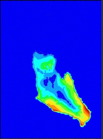
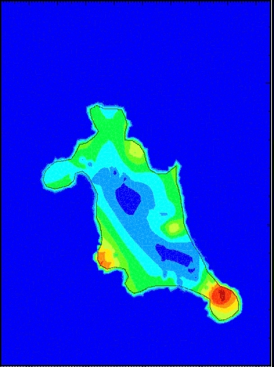
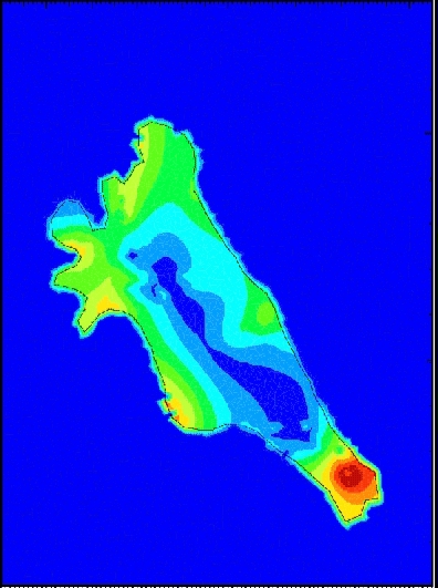
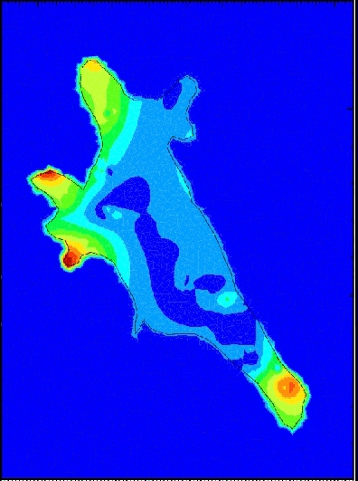
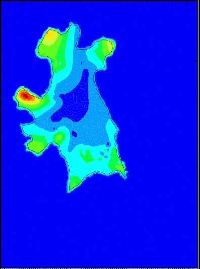
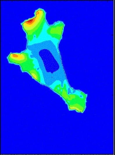
Fig. 5 Traction forces exerted by a T24 cancer cell on a rigid PolyAcrylamide substrate (E=10kPa)
see also
Book "Cell mechanics: from single scale-based models to multiscale modeling", Eds. Arnaud Chauviere, Luigi Preziosi, Claude Verdier
CRC Press/Chapman & Hall, january 2010
Viscoelastic properties of tissues (micro-macro scales)
The mechanical properties of a model tissue depend largely on the ability of cells to deform the matrix, i.e. collagen for example within connective tissues.
We have shown that some cells pull on the collagen fibers and remodel collagen (Fig. 6):
1. By attracting fibers near them
2. By bringing along fibers as they migrate
These two effects can have a dramatic effect on the tissue resistance (breakdown of the elastic network)
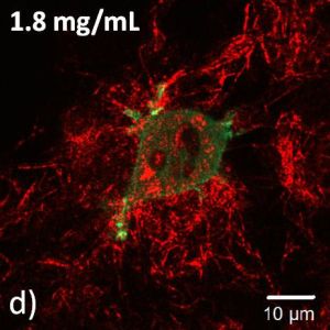
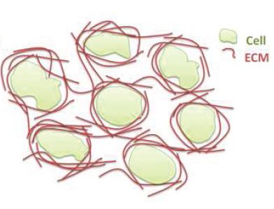
Fig. 6 CHO cells embedded in a collagen matrix. Cells are labeled in green with GFP (left), collagen fibers are seen in red thanks to the
reflectance technique on a confocal microscope. Right: sketch of the remodelling process
Spheroids rheology
Spheroids are produced in the lab and their global rheology is obtained using large cantilevers (50µm width). A special AFM tool is used to measure microrheology after an initial indentation (Fig. 7)
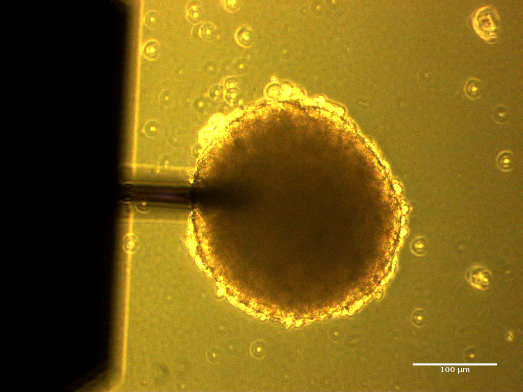
Fig. 7 Sketch of the experimental technique. (a) Spheroid indentation (scale 200µm). (b) Sinusoidal small deformations. (c) Viscoelastic data for T24 spheroids with added collagen (content from 0 to 0.03 mg/mL)